The Different Biopsy Types
Different types of biopsies can be taken depending on a few factors, such as lesion type and location. Your dermatologist will recommend which option is most suitable for the area of your body needing sampling while making the impact on you and your lifestyle as minimal as possible. If you have a preference for the type of skin biopsy you would like, you are more than welcome to let your dermatologist know about this.
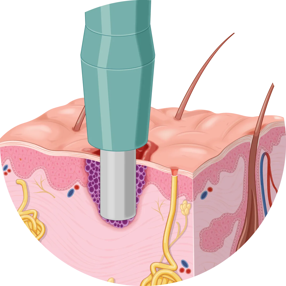
Punch Biopsy
A punch biopsy procedure involves a tool that takes a cylindrical piece of skin followed by stitches to close the wound. You will generally need to return to the clinic approximately one week later to have these stitches removed. The main advantage of a punch biopsy procedure is that it can sample deeper layers of skin. However, you must look after the wound to minimise the risk of the stitches coming apart.
Your dermatologist may end up removing the whole lesion from the sample for very small lesions. This is sometimes referred to as a punch excision.

Shave Biopsy
A shave biopsy involves taking a superficial shave of skin to confirm the diagnosis. It is used when a deeper assessment of the lesion is unnecessary. The main advantage is that stitches are not usually required because the tissue sample is taken superficially, so you won’t need to return to the clinic. The wound heals like a graze and has a minimal impact on your normal activities. In many cases, a shave wound heals without scarring.
Curettage (or Scrape) Biopsies
A curettage is very similar to a shave; however, a different removal instrument called a “curette” is used. Like a shave, the main advantage of curettage is that it only removes superficial tissue and closing the wound with stitches is generally not required. It heals like a graze and is particularly useful in concave areas where it is difficult to use a shave biopsy blade.

Incisional Biopsies
An incisional biopsy takes a thin, deep, and long sliver of a lesion to sample the entire cross-section of a lesion. It is beneficial for pigmented lesions in cosmetically sensitive or difficult areas. This provides a reliable assessment while avoiding the full removal of the lesion in case this later proves unnecessary. An incisional biopsy can be thought of as a mini-excision.
Ordinary stitches will be placed to close the wound. This requires you to look after the wound, and you will need to return to the clinic about a week later to have the stitches removed.
