Dermoscopic Features
Some of the features that your dermatologist will be looking for with a dermatoscope include:
- The substructure of the lesion, the pigment network, and how the pigment is organised and distributed across the lesion. This may include lines, dots, clods, and areas without structure or pigment.
- Whether the substructure is symmetrical or asymmetrical and the extent of how chaotic or disordered the lesion is.
- The presence of blood vessels, telangiectasia, and their shape and distribution.
- Other features such as keratin (scale), cysts, and fissures.
Dermoscopy at Skintel
This simple appointment is an in-depth evaluation of your skin that’s carried out by a specialist dermatologist to ensure you have the most thorough examination available for skin cancer detection. Our specialists provide the utmost professionalism and to make you feel comfortable, we also provide a specialised gown you can wear during your examination.
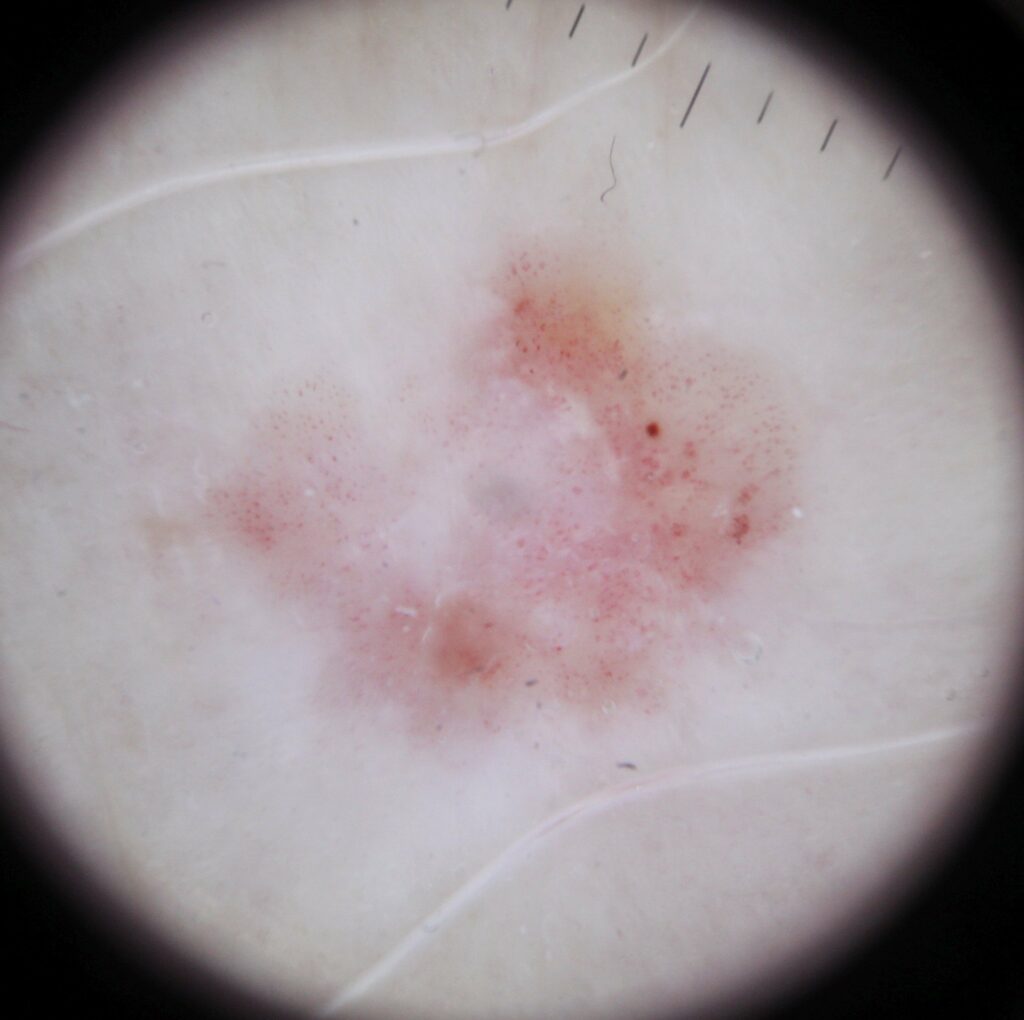
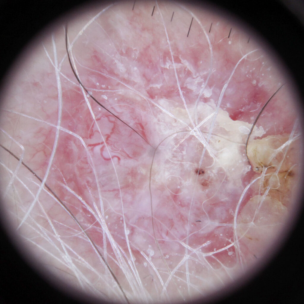
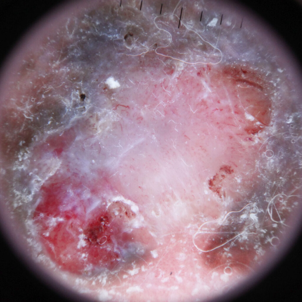
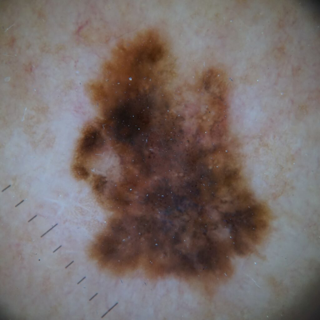
Dermoscopic view of common skin cancers

Frequently Asked Questions
Is dermoscopy painful?
Having a skin cancer dermoscopy by a dermatologist is not a painful procedure at all. They may apply some very low-level pressure when using the dermatoscope on your skin.
Is dermoscopy safe?
Having a Dermoscopy is a very safe process. It is a simple procedure that involves a dermatologist looking over your skin surface for suspicious patches of skin or other signs of melanomas with a handheld magnifying glass.
How long does a dermoscopy take?
Generally, a skin cancer dermoscopy takes anywhere up to 30 minutes. It all depends on your body. If you have many concerning lesions, they may need some extra assessment time.
What does skin cancer look like under a dermatoscope?
There are many different aspects of how a skin cancer can look under a dermatoscope. Some of these include irregularity in blotches, spots, streaks, regression structures, atypical networks, and possibly atypical vascular patterns. A dermatologist is the best specialist to see for this basic procedure. It must be noted that to diagnose skin cancer, a biopsy must be taken.


